Thinking about something, darling?
We would often like to be able to look inside another person’s mind. Thomas Beyer from the Medical University of Vienna is developing methods that are able to do just that. The technique he and his team use combines the well-known magnetic resonance imaging (MRI), which takes 3D images of soft tissue, with the lesser-known positron emission tomography (PET), which can determine the brain’s sugar metabolism. The aim is to produce something like a map of brain activity. In a project funded by the Austrian Science Fund FWF, his team has now developed an automated procedure which has major advantages over the methods in current use.
Blood sampling required
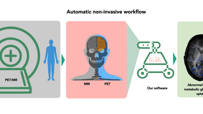
Usually, an accurate measurement of the brain’s metabolism is very unpleasant, explains Beyer: “Arterial blood is sampled from the wrist, and the test persons have to lie still for one hour.” Beyer's hypothesis is that a PET/MRI scanner could be used to avoid taking blood samples. Such a device is capable of combining MRI images, which make soft tissue contrasts visible, with PET images. For the PET scan, the test persons are administered a small amount of radioactively labelled sugar. “One injects the radioactive glucose into the bloodstream where it gets distributed. The sugar we use for this procedure is labelled with the radioactive element fluorine 18, which emits two photons after decay,” Beyer explains. These photons can be detected with the PET scanner and the resultant image shows where the sugar is processed in the brain. The MR image serves as a reference because people in the scanner might not lie entirely still, and this could blur the PET scans. According to Beyer, the resulting exposure to radioactivity is within the range of the standard annual amount of radiation a person in Austria is exposed to through natural influences. PET/MRI scanners are still very rare, says Beyer. “In the whole world there are about 250 of them, in Austria there is just one, and that’s the one at the Medical University of Vienna.” Beyer emphasises that the images generated with PET/MRI are more than just a superimposition of two individual images: “We use information from the MRI to make the PET images better and more accurate.”
Brain activity without blood sampling
In the clinical project, the researchers examined a group of ten test persons with this device in collaboration with partners from the Medical University of Vienna. After being administered the contrast medium, radioactive sugar, the test persons lay in the PET/MRI scanner for 60 minutes, during which time blood was sampled regularly. “This was necessary to see whether what we measure in the brain corresponds to the blood glucose content,” says Beyer. The comparison with the classical method by blood sampling confirmed the reliability of PET/MRI measurements of brain activity, which means that it will be possible to forego the blood sampling for this examination in the future. This will enable physicians to use quantitative measurements of brain metabolism in clinical practice.
Application for epilepsy
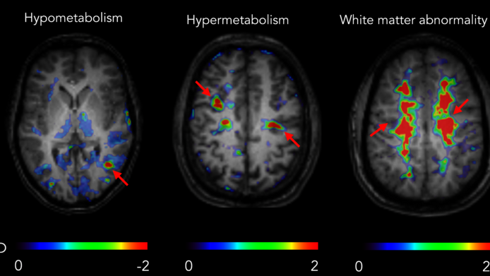
After this first successful study, the researchers investigated whether the method could be used in support of epilepsy treatment. In non-lesional epilepsy – a form of the disease that is not caused by anatomical factors – it is crucial to know the activity of certain brain regions, which are then the target of an intervention in which the problematic tissue is surgically removed. In order to ascertain which region is responsible for the epileptic seizures, precise measurements are needed to determine that region’s activity. The sugar metabolism of such an area is low when the patient is at rest, but rises sharply during an epileptic seizure. “The sugar metabolism is not the best indicator for epileptic foci, but we wanted to find out whether it was suitable for diagnosing them,” said Beyer. “We therefore chose not only to take a static picture after a certain point in time when the labelled sugar is evenly distributed, but also to document its accumulation over time.” This enables the research team to see how “hungry” the brain cells are. “We hoped this approach might enable us better to describe the epileptic zones.”
However, in a first study involving 15 patients with non-lesional epilepsy, the hopes of the researchers were not fulfilled. The PET/MRI imaging procedures and the quantification they developed provided some added value only in a small fraction of the people examined. “This can be explained by the fact that the baseline sugar consumption also fluctuates strongly in a normal brain,” says Beyer. “It depends on the patient’s state of mind, which is subject to significant fluctuations – the brain’s sugar consumption changes depending on whether one is in a good mood or stressed. So far, however, we have not been able to standardise the patient's state of mind, which remains a challenge for the future.” The researchers were unable to distinguish the disease-related changes in sugar metabolism from these natural fluctuations.
Automated determination of brain activity
Accordingly, the Viennese research team has focused more on general non-invasive measurements of brain activity in healthy people. This approach proved to be successful. Thanks to the work of Beyer's team member Lalith Kumar Shiyam Sundar, a biomedical engineer who acquired his doctorate in the course of the four-year project, it was possible to automate the examination to a large degree. The computer now reconstructs brain activity from the image data. Recently, the quality of the images has been further improved by means of machine learning methods. “The methodology we have developed is automated so that physicians can use it for neuro-oncological examinations, for instance,” notes Thomas Beyer. “For these, we have found a solution here, which displays this new type of images at the push of a button.”
Personal details Thomas Beyer is a physicist at the Medical University of Vienna and Head of the Quantitative Imaging and Medical Physics research unit. He is President of the European Society for Hybrid, Molecular and Translational Imaging and a member of the European Academy of Sciences. Beyer is particularly interested in hybrid imaging methods in medicine and their application in areas such as neurology and oncology. The clinical basic-research project “Personalised diagnosis of non-lesional epilepsy using simultaneous PET/MR imaging” (2015-2019) was supported by the Austrian Science Fund FWF with an amount of approximately 245,000 euros.
Publications
