Gabriele Colozza
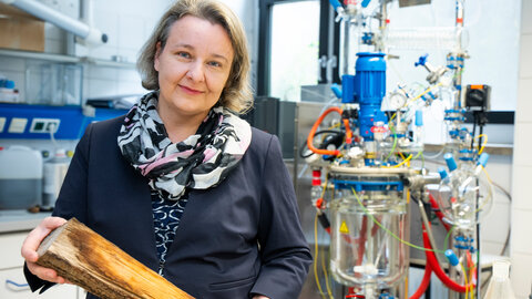
Gabriele Colozza succeeded in identifying a gene that illustrates how stem cells in the intestine make decisions. His discovery is an important step towards advancing therapies for colorectal cancer in a targeted way. © Hanna Gabriel/FWF

Stem cells vary in their degree of potency. Totipotent stem cells can develop (or “differentiate”) to form all types of cells in an organism. Probably the best-known example is the fertilised egg cell, which divides to form the entire body with all its different types of tissue. In contrast, adult stem cells are multipotent. They are present in fully formed tissue and specialise in generating certain types of cells. Intestinal stem cells, for instance, can only form the cell types of intestinal mucosal membranes.

“Adult stem cells are important because they regenerate dead or damaged cells in tissue in order to ensure its functionality,” says Gabriele Colozza. The Italian-born scientist currently holds a research fellowship at the Institute of Molecular Biotechnology (IMBA) in Vienna, funded by the Lise Meitner Programme of the Austrian Science Fund FWF to promote international mobility. In his research project on molecular mechanisms regulating adult intestinal stem cells, he has identified a gene that plays an essential role in the differentiation of intestinal stem cells. It acts as a switch for the formation of so-called Paneth cells, a type of secretory cell in the intestines. Certain types of intestinal cancer are characterised by excessive proliferation of Paneth cells, which is why scientists see them as a new therapeutic target at the cellular level.

Intestines constantly under construction

“The cells in our intestines are exposed to harsh conditions,” says Colozza. They experience mechanical wear and tear due to the passage of food and are also confronted with digestive enzymes and varying pH values. “Whether cells die of natural causes or due to external factors, you need a potent force to regenerate intestinal mucosal membranes,” explains the researcher. That is the function of intestinal stem cells. Mouse intestines have to completely regenerate every three to five days, according to estimates. The human intestine should similarly be seen as a dynamic balance rather than a set structure, says Colozza.

Stem cells supply replacements

The intestinal mucosa’s characteristic structure features protrusions about 1 millimetre long, called intestinal villi. They enlarge its surface and are crucial in the absorption of nutrients. Located in the interstices between the villi are so-called crypts, small pocket-like structures. “In each crypt, there is a multitude of stem cells that can differentiate to form intestinal cells,” explains Colozza. “What interests me is what molecular signals tell a stem cell which path to take in its differentiation.” The further development of stem cells is regulated by their environment.

Unchecked signalling pathways lead to cancer

“Cells use molecular signals for communication,” explains Colozza. One of these signalling pathways is called Wnt. It plays a significant role in embryonic development and can lead to excessive cell division and consequently to cancer if left to act unchecked.

One of the best-known antagonists of the Wnt signalling pathway is Rnf43. The gene was identified by Colozza’s original laboratory supervisor in Vienna, Bon-Kyoung Koo. It is essential for getting rid of proteins that are no longer needed, marking these inside the cell when they need to be eliminated – among them the receptor of the Wnt signalling pathway. By this, Rnf43 dampens the Wnt signal to the extent necessary. To do so, however, it needs the right partner protein.

Personal details

Gabriele Colozza is a Senior Scientist at the Institute of Molecular Biotechnology (IMBA) in Vienna. After completing his degree in Siena, he pursued research in Cambridge (United Kingdom) as well as in the USA at the University of California, Los Angeles, and Columbia University in New York.

Since 2020, he has worked in Vienna as a postdoc fellow. His project on molecular mechanisms regulating adult intestinal stem cells is funded by a Lise Meitner grant of the FWF in the amount of EUR 175,780.

Fluorescent immunostaining of the mouse intestine.
Fluorescent immunostaining of the mouse intestine. The crypts (invaginations or pocket-like structures) are located between the intestinal villi (long, finger-like structures that extend into the intestinal lumen). Paneth cells, a type of glandular cell found in the crypts, are coloured green. Blue colouring shows the nuclei of all intestinal cell types. © Gabriele Colozza

Molecular teamwork decoded

Earlier research had shown that marking by Rnf43 in itself was not enough to break down the Wnt receptor. Like most receptors, the Wnt receptor is located in the membrane surrounding the cell, its plasma membrane. For marking by Rnf43 to lead to its breakdown, the receptor would first have to be absorbed into the cell. “This is where our research project comes in,” says Colozza. “Using biochemical assays and mass spectrometry, we were able to analyse which proteins interact with Rnf43.” Colozza identified a gene called Daam as the missing link. “It ensures that the Wnt receptor is incorporated into the cell. This means that Daam and Rnf43 work in tandem to dampen the Wnt signalling pathway,” the expert explains.

Effects investigated in intestinal organoids

To translate the discovery from the molecular level to the organ level, one of the methods Colozza used were intestinal organoids. These three-dimensional structures are grown as cell cultures from adult stem cells and have a structure and properties similar to those of intestinal mucosa. Colozza used them to determine that Daam is essential for the differentiation of intestinal stem cells to form so-called Paneth cells. These cells secrete substances that stimulate cell division in intestinal stem cells. “Daam acts as a switch,” the scientist explains. “When it is switched on, the stem cell differentiates to form a Paneth cell. When it is switched off, the cell takes another path.”

More Paneth cells in colorectal cancer

The link between the results at the molecular level and the development of Paneth cells lends the project additional importance. This is because one of the functions of Paneth cells is to regulate ambient conditions in the crypts. Since Paneth cells stimulate stem cell division, their excessive prevalence can contribute to the development of cancer.

“Mouse models have been used to show that such tumours recede when the excessive proliferation of Paneth cells is genetically blocked,” Colozza explains. “Our research has delivered the first genetic proof that a component of the Wnt signalling pathway is directly involved in the development of this crucial type of cell.” This also puts a new goal within reach in the development of drugs against colorectal cancer.

Results enhance understanding of stem cells

“Our results demonstrate how stem cells make decisions,” says Colozza. That is a boon to science as such in his opinion. “While we know how to cultivate certain cells in the lab, we often lack the means to push them in a specific direction.” This is changing with increasing insight into genetic and molecular mechanisms. The identification of Daam is an important step towards being able to specifically stimulate the development of stem cells into particular types of cells.